


Retinal Disease Treatment
Retinal disease treatment refers to the medical care used to prevent, manage, or treat diseases and conditions affecting the retina, the light-sensitive layer of tissue at the back of the eye.
RETINA TEAR
The retina is a transparent membrane about 0.2 mm thick, located on the innermost layer of the eyeball. It is composed of 10 layers and, in camera terms, plays the role of film. External information enters through the cornea, passes through the lens and vitreous humor, and reaches the retina. The retina contains photoreceptor cells that sense light, color, and shape, and plays a crucial role in converting light information into electrical signals. These converted electrical signals are then transmitted to the brain via the optic nerve, which runs throughout the retina, allowing us to see.
WHAT IS RETINA?
A retinal tear is a tear in the retina, and if left untreated, it can lead to retinal detachment. Retinal tears are classified into “atrophic tears,” which occur due to degeneration and atrophy of the vitreous humor, and “tractional tears,” which occur when the retina is pulled by the vitreous humor due to adhesion between the vitreous humor and the retina. Of these, “tractional tears” are particularly prone to leading to retinal detachment.
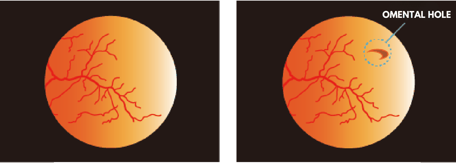
RETINAL TEAR CAUSED BY POSTERIOR VITREOUS
DETACHMENT DUE TO AGING
The inside of the eyeball is filled with a jelly-like substance called the vitreous humor, but with age, the vitreous humor changes into a thinner, more liquid-like substance.
This is called vitreous degeneration. When vitreous degeneration occurs, the volume of the vitreous humor decreases, and posterior vitreous detachment occurs, where the vitreous humor separates from the posterior part of the retina.
At that time, holes or tears may form in weaker parts of the retina or in areas where the vitreous humor and retina are strongly adhered.
RETINAL TEARS CAUSED BY HIGH MYOPIA
Myopia occurs when the length (depth) of the eyeball increases, causing the focal point to be in front of the retina. As the depth of the eyeball increases, the retina is stretched to accommodate this elongation, which can create thin and weak areas. When these areas atrophy, they can develop into retinal tears.
RETINAL TEAR CAUSED BY CONTUSION TO THE EYEBALL
Severe impacts to the head or eyes, such as those caused by strenuous exercise or falls, can rapidly alter the eyeball, potentially leading to retinal tears.
The main symptoms of a retinal tear include “floaters,” which are black spots or specks seen in the field of vision, and “photopsia,” which is the perception of light even when there is no light. Particular attention should be paid if there is a sudden change in what you see. If the retinal tear progresses and causes retinal detachment, it can lead to decreased vision or visual field defects, and if left untreated, it can lead to blindness.

NOTE: ACTUAL APPEARANCE MAY VARY FROM THE PERSON AND MAY DIFFER FROM THE IMAGE ABOVE.
Common symptoms of retinal tears include “floaters” and “photopsia.” Floaters, where you see black spots or specks in your field of vision, and photopsia, where you see flashes of light even when there is no light, can both be caused by vitreous opacities resulting from aging or nearsightedness.
However, you should pay particular attention if your vision suddenly changes. If a retinal tear is left untreated, fluid from the vitreous humor can enter through the hole, causing retinal detachment. Retinal detachment can lead to decreased vision and visual field defects, and if left untreated, it can result in blindness, making it a very dangerous eye disease.
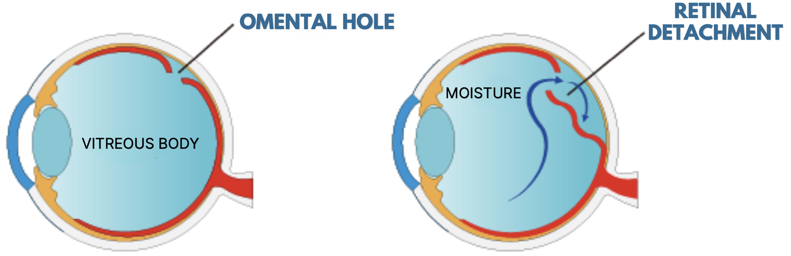
If a retinal tear is left untreated, fluid from the vitreous humor can enter through the hole, leading to retinal detachment.
The examination procedure involves a visual acuity test followed by a detailed examination of the fundus. The fundus examination is a detailed examination of the retina and is performed after administering pupil-dilating eye drops.
After pupil dilation, you will feel sensitive to light for about 3 to 6 hours, so you will not be able to drive a car immediately after the examination, but your eyes will return to normal naturally over time.
In addition, examinations using OCT (Optical Coherence Tomography), a fundus examination device that uses near-infrared light, allow us to observe cross-sections of the retina that were previously impossible to observe. This examination has made it possible to make a precise diagnosis of retinal diseases.
OCT (OPTICAL COHERENCE TOMOGRAPHY)
EXAMINATION
OCT (Optical Coherence Tomography) is a state-of-the-art diagnostic tool that can capture cross-sectional images of the retina, obtaining images with a precision close to that of an optical microscope. By shining near-infrared light onto the fundus of the eye and analyzing the reflected waves, it is possible to examine the cross-section of the retina.
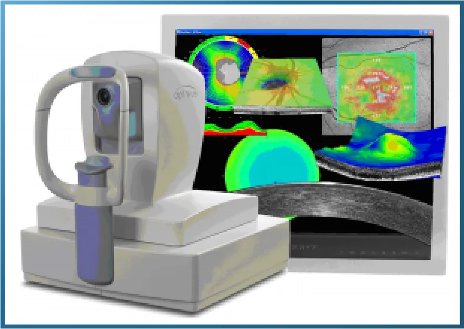
While conventional examinations could only observe the retina in a two-dimensional manner, the advent of OCT makes it possible to observe the retina in cross-section, allowing for examination of retinal edema, the extent and depth of bleeding, and even the condition of the optic nerve.
Because there are few facilities that have introduced OCT, the number of facilities where you can receive this examination is limited. However, it is very useful for diagnosing retinal diseases and glaucoma, and allows for accurate diagnosis of treatment plans, treatment effectiveness, and treatment progress.
One treatment method for retinal tears is laser photocoagulation, which involves using a laser to seal the area around the tear. When fluid from the vitreous humor enters the area beneath the retina through a tear, the tear widens and progresses to retinal detachment.
To prevent progression to retinal detachment, the area around the tear is sealed with a laser to prevent fluid from entering. If a retinal tear is discovered during an examination, our clinic strives to provide prompt treatment with the patient’s consent.
The retina can detach in as little as one day, so early detection and treatment are crucial to prevent progression to retinal detachment. Retinal tears have a higher risk of leading to retinal detachment than retinal holes, so early treatment is necessary.
HIGH-PERFORMANCE LASER
INTRODUCING THE PASCAL SYNTHESIS OPHTHALMIC
LASER PHOTOCOAGULATION DEVICE.
We have introduced the “PASCAL Synthesis” ophthalmic laser photocoagulation system. This laser system ensures exceptional precision and high visibility, and is suitable for a variety of treatments including retinal photocoagulation, posterior capsule opacification treatment, glaucoma treatment, and iridotomy. Its unique beam profile and sharp irradiation spot enable safe treatment without affecting the cornea, and by focusing the laser energy to the target area to the maximum extent, it minimizes the energy required for treatment.
Equipped with a high-performance combination device that allows switching to the appropriate laser treatment for the treatment purpose with the push of a button, it is designed for use in limited spaces. The laser system is compactly designed with excellent design, ensuring sufficient space for treatment, and its control panel operation enables comfortable operation and accurate treatment.
HIGH-PERFORMANCE LASERS
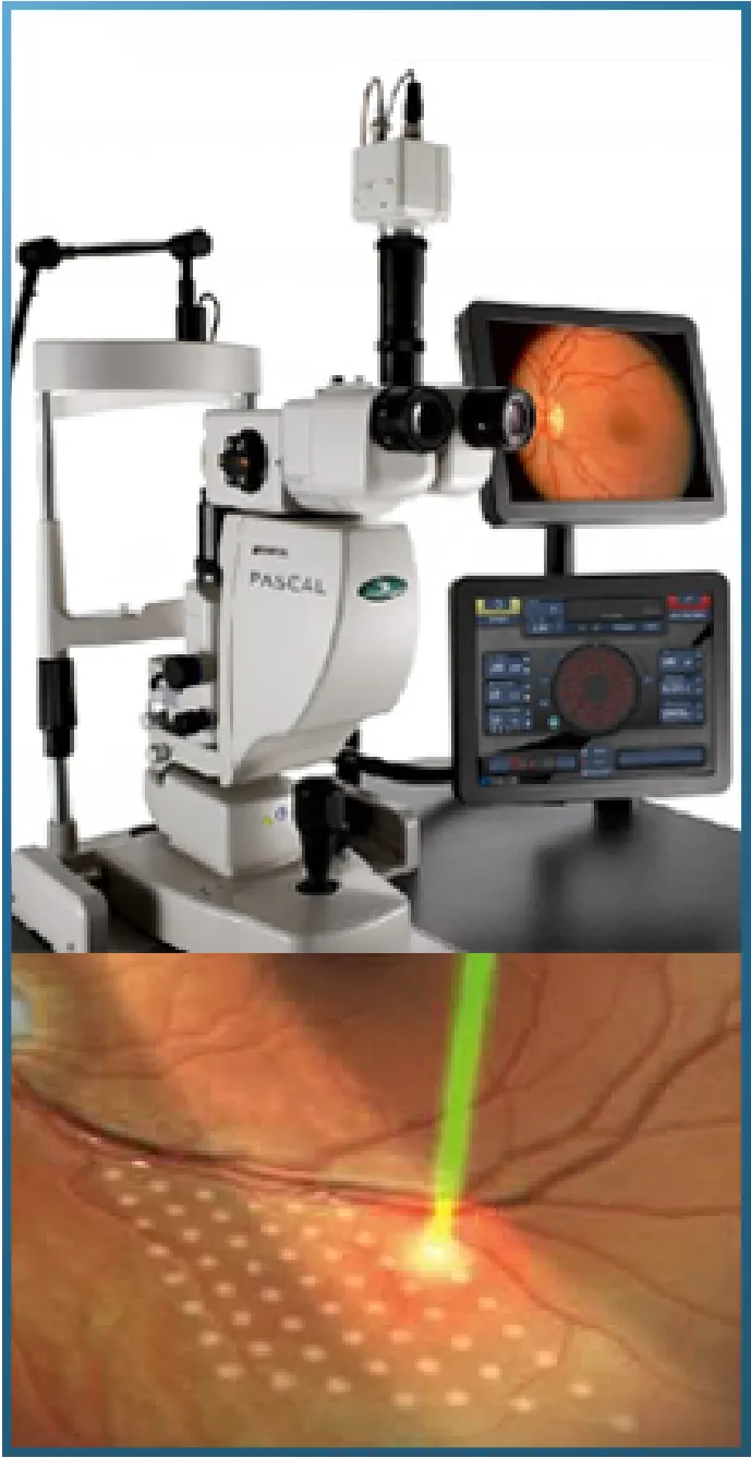
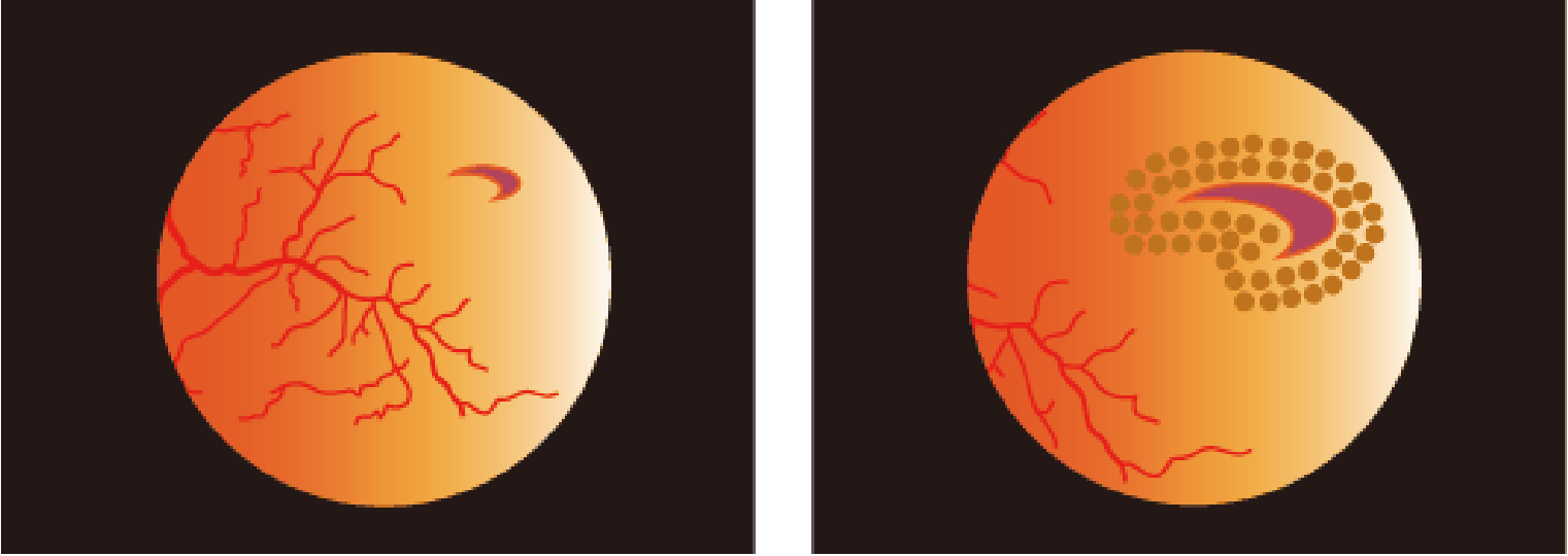
SEALED WITH A LASER
WE RECOMMEND REGULAR CHECKUPS FOR RETINAL
TEARS, WHICH CAN LEAD TO BLINDNESS.
Retinal tears are commonly seen in people with severe myopia and in middle-aged and older adults. If left untreated, they can progress to retinal detachment, leading to decreased vision, visual field defects, and in the worst cases, blindness. Therefore, it is recommended that people with severe myopia and those over 50 years old undergo regular retinal examinations.
RETINAL
DETACHMENT
The retina is a transparent membrane about 0.2 mm thick, located on the innermost layer of the eyeball. It is composed of 10 layers and, in camera terms, plays the role of film. External information enters through the cornea, passes through the lens and vitreous humor, and reaches the retina. The retina contains photoreceptor cells that sense light, color, and shape, and plays a crucial role in converting light information into electrical signals. These converted electrical signals are then transmitted to the brain via the optic nerve, which runs throughout the retina, allowing us to see.
Retinal detachment is a condition in which the retina, a membrane located on the inside of the eyeball, detaches, leading to decreased vision. The retina is the tissue that receives light entering the eye as stimuli and transmits them to the optic nerve in the brain; it functions like the film in a camera.
Retinal detachment is often painless and therefore difficult to notice, but floaters (vitreous opacities) may appear as a warning sign. Furthermore, if the detachment extends to the macula, the central part of the retina, vision can deteriorate rapidly, potentially leading to blindness.
Retinal detachment can be caused by aging, certain diseases such as diabetic retinopathy, or physical shock to the head or eyeball due to an accident. In all cases, a tear in the retina (retinal tear) is the first step in retinal detachment.
The inside of the eyeball is filled with a gel-like substance called the vitreous humor, but sometimes a part of the retina can be dragged into this vitreous humor, creating a small tear in the retina. If the tear is left untreated, fluid will gradually seep into the space between the retina and the layer beneath it, eventually causing the retina to peel off completely.
IF THERE IS A TEAR
One treatment to close tears in the retina is “photocoagulation.” This involves shining a laser through the pupil into the hole in the retina and cauterizing it. This procedure causes the retina surrounding the tear and the tissue beneath it to stick together, making it less likely for the retina to detach.
IF THE RETINA IS DETACHED
If retinal detachment is already present, it is necessary to fix the detached retina back into place. Surgical procedures for this include “vitrectomy” and “scleral buckling,” and the appropriate treatment varies depending on the severity of the retinal detachment.
Moving your eyes after surgery will not significantly affect the outcome, but it takes 1 to 3 months for the inside of the eye to stabilize. For at least one month after surgery, use your eyes to the extent that you do not strain them.
Those who do desk work such as office work or management can return to work from one month after surgery, while those who do physically demanding work such as driving can return to work from around two months. In daily life, avoid lifting heavy objects, strenuous exercise such as running, and driving a car for one month after surgery.
The earlier retinal detachment is treated, the less impact it has on vision, so early detection and prompt treatment are crucial. If you are concerned about any of the following symptoms, please have your eyes examined by an ophthalmologist.
– I see small, black, mosquito-like objects moving around in front of my eyes.
– I suddenly see flashes of lightning in dark places.
– My eyesight has suddenly deteriorated.