


Glaucoma Treatment
Glaucoma is a disease in which abnormalities occur in the optic nerve, preventing accurate transmission of information from the eyes to the brain. As a result, the brain is unable to properly recognize the information as an image, leading to impaired vision and visual field (the area that can be seen). There are several types of glaucoma, but as the disease progresses, a symptom called “visual field defect” appears, in which the blind spots in the visual field gradually increase.
EARLY DETECTION AND TREATMENT
ARE CRUCIAL FOR GLAUCOMA
As with all illnesses, early detection and treatment are crucial for eye diseases. Eye diseases, in particular, are often overlooked because they have progressed significantly by the time symptoms become noticeable.
Glaucoma, in particular, does not heal on its own once it has progressed.
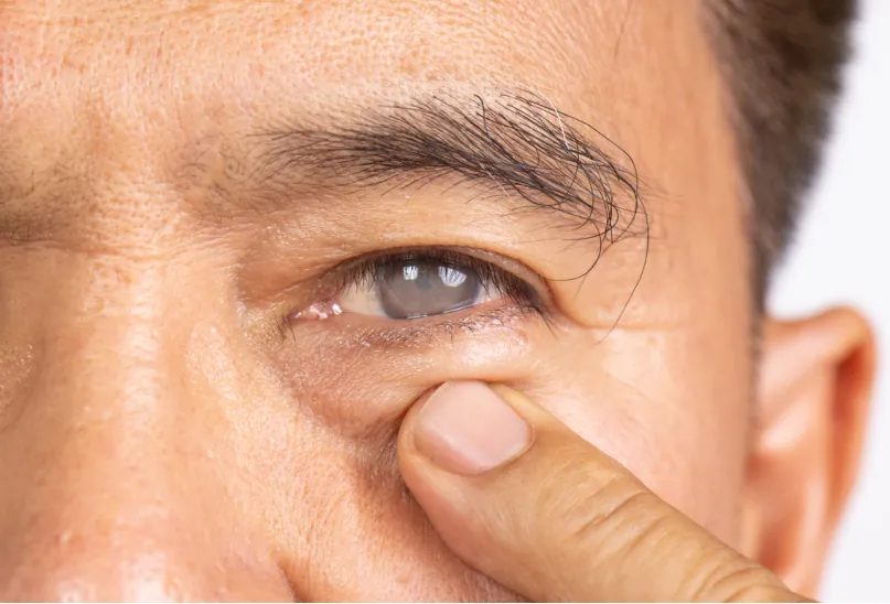
If you experience any abnormalities such as blurred vision or hazy eyesight, we recommend consulting an ophthalmologist. This will lead to early detection and treatment.
MAIN SYMPTOMS THAT INDICATE EYE PROBLEMS
- Blurred vision/blurred vision
- Itchy eyes/eye pain
- Excessive eye discharge
- Redness/swelling of the eyes
- Decreased vision
- Distorted vision
- Loss in the field of vision
Glaucoma is a disease in which the optic nerve is compressed due to elevated intraocular pressure caused by various factors, resulting in damage to a portion of the compressed optic nerve. The optic nerve, located within the retina, is bent in bundles at the optic nerve head, but the bends are very weak.
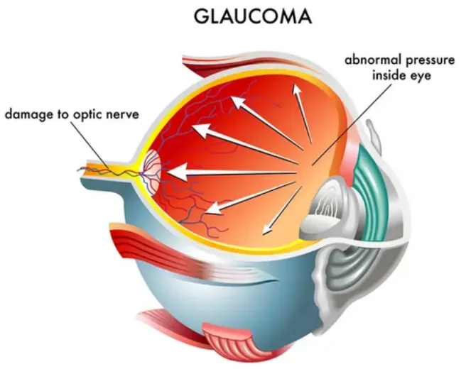
When some of the nerve fibers are damaged due to increased intraocular pressure, the information entering the eye cannot be accurately transmitted to the brain, resulting in symptoms of partial vision loss (visual field defects). Once damaged, nerve fibers do not regenerate, so it is impossible to recover lost vision.
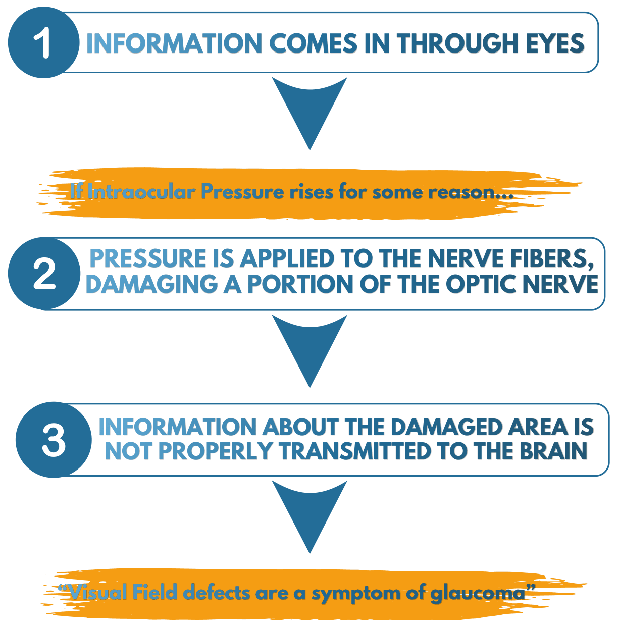
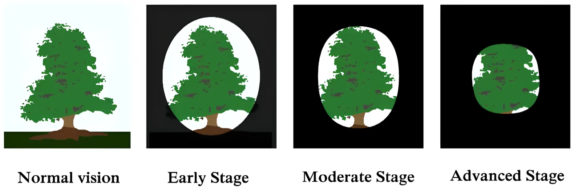
If the disease progresses, it may lead to blindness.
WHAT IS “INTRAOCULAR PRESSURE” WHICH
CLOSELY RELATED TO GLAUCOMA?
Intraocular pressure plays a significant role in glaucoma, as it plays a crucial role in maintaining the round shape of the eyeball. The outermost layer of the eyeball is a very strong membrane called the sclera. This is the part commonly known as the white of the eye, and within it is a jelly-like tissue called the vitreous humor, which makes up the majority of the eyeball. In front of the sclera are the cornea, a transparent membrane, and the lens, which acts as a lens.
However, these tissues alone cannot maintain the round shape of the eyeball. A clear fluid called aqueous humor plays a major role in maintaining the round shape of the eyeball, and it primarily performs two roles.
First, it transports nutrients into the eye and removes waste products. Second, it regulates intraocular pressure. Just as a ball becomes harder when filled with air, the firmness of the eyeball is determined by the amount of aqueous humor. The balance between the production and drainage of aqueous humor allows the eyeball to maintain its spherical shape through internal pressure. The normal intraocular pressure (IOP) for Japanese people is 10-20 mmHg, with an average of 14.5 mmHg. However, IOP can fluctuate when the balance between aqueous humor secretion and drainage is disrupted. An increase in the amount of aqueous humor inside the eye raises IOP and can cause glaucoma.
1.Nutrient transport and waste removal
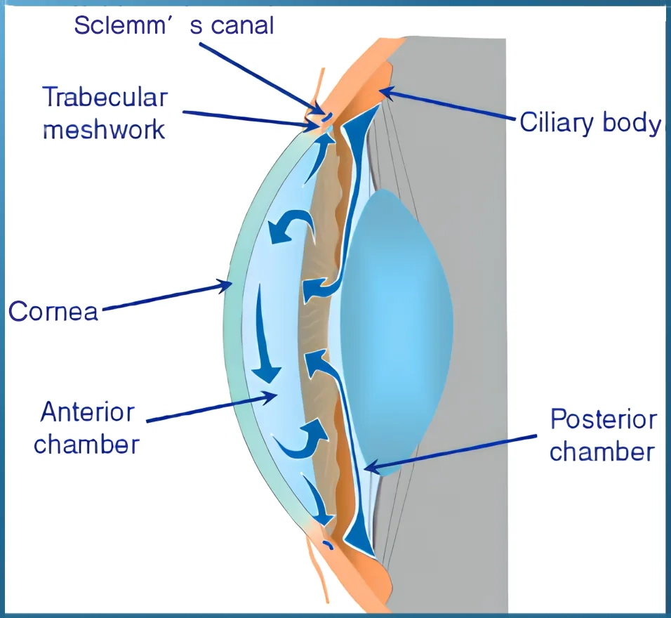
- Aqueous humor is produced in the ciliary body and plays a role in transporting nutrients into the eyeball and draining waste products through Schlemm’s canal.
2.Maintain normal intraocular pressure
- The amount of aqueous humor inside the eye affects the firmness of the eyeball. Inside the eye, the secretion and drainage of aqueous humor are controlled to remain constant. This creates a constant pressure from the inside of the eyeball, maintaining its round shape.
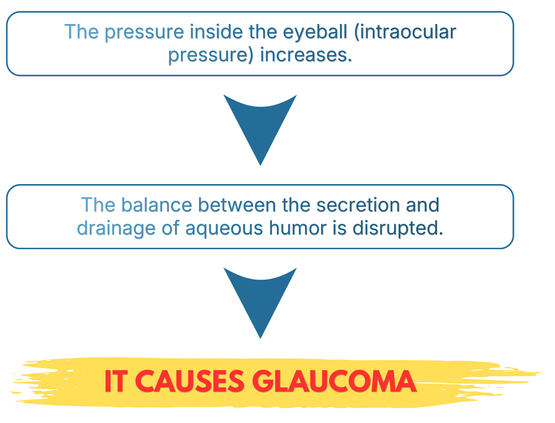
Glaucoma can be categorized into several types, each with different causes and symptoms. Broadly speaking, there are two main types: glaucoma caused by elevated intraocular pressure and glaucoma that does not involve elevated intraocular pressure. Other types include congenital glaucoma and glaucoma caused by eye trauma or other diseases. Treatment methods vary depending on the cause and symptoms, so it is important to first undergo examinations to determine the cause.
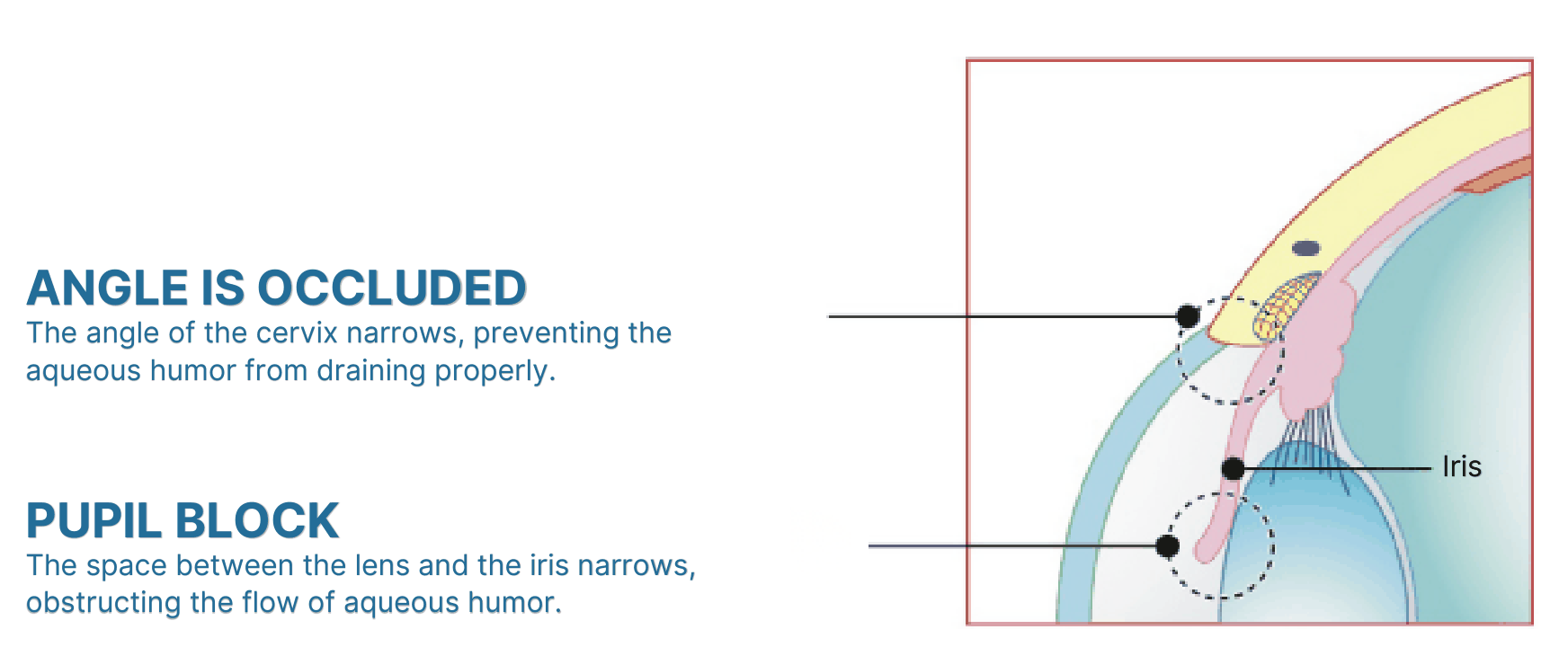
Inside the eye, a fluid called aqueous humor is constantly produced and drained, maintaining intraocular pressure while simultaneously supplying nutrients to the cornea and lens. Aqueous humor is produced in the ciliary body and drained through Schlemm’s canal. Between the cornea and the iris is an area called the iridocorneal angle, through which aqueous humor flows. If the iridocorneal angle is blocked by the iris, the produced aqueous humor loses its drainage route, causing intraocular pressure to rise.
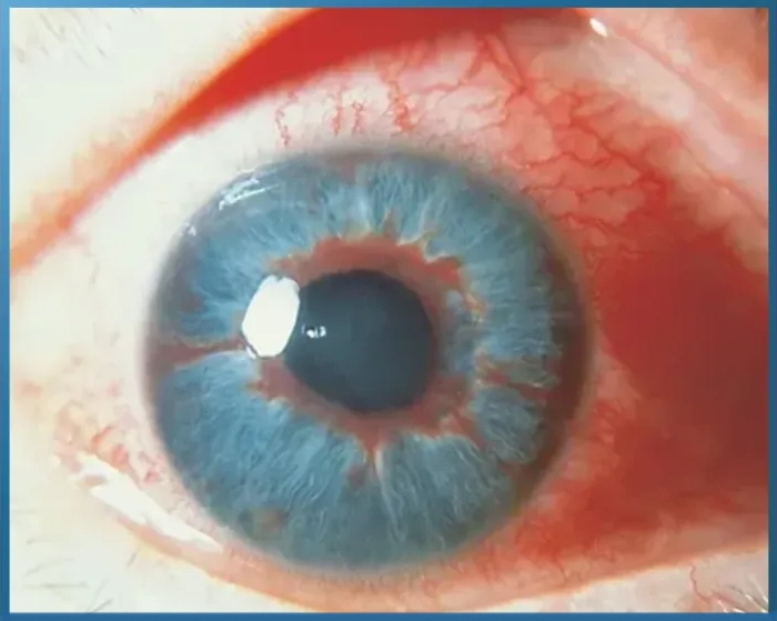
Glaucoma caused by this is called primary angle-closure glaucoma. Primary angle-closure glaucoma is characterized by a rapid increase in intraocular pressure because the flow of aqueous humor is completely stopped, and is also called “acute glaucoma.” Because symptoms appear suddenly and progress rapidly, blindness can occur if appropriate treatment is not given within 48 hours of the onset, making it one of the most dangerous eye diseases. Primary angle-closure glaucoma is more common in women than men, and its prevalence increases with age.
PRIMARY ANGLE-CLOSURE GLAUCOMA IS
MORE LIKELY TO OCCUR IN THESE SITUATIONS:
- When looking downwards for a long time
- When excited
- When straining your eyes too much
- When looking at things in a dark place
- Insomnia
- overwork
- excessive stress,
- or after using pupil-dilating eye drops.
MAIN SYMPTOMS OF PRIMARY ANGLE-
CLOSURE GLAUCOMA
- Severe eye pain
- blurred vision, decreased visual acuity
- Iris vision (seeing rainbows around lights) and
headaches. - Nausea
- Red eyes
- Corneal edema
- Corneal opacity
- Pupil dilation
Aqueous humor passes through a mesh-like organ called the “trabecular meshwork,” which acts as a filter, and is then drained through Schlemm’s canal. If this trabecular meshwork becomes clogged with cellular debris contained in the aqueous humor, the aqueous humor cannot be properly drained, and intraocular pressure rises.
Glaucoma caused by this is called primary open-angle glaucoma. Primary open-angle glaucoma is said to be caused by genetics, but it is more commonly seen in people in their 40s and 50s who are nearsighted or have diabetes. The frightening thing about this disease is that it often goes unnoticed because the only symptoms are headaches and eye strain. The disease progresses over many years, making it difficult to notice symptoms such as visual field defects.
PRIMARY OPEN-ANGLE GLAUCOMA IS MORE LIKELY TO
OCCUR IN THE FOLLOWING TYPES OF PEOPLE:
- If you have a family history of glaucoma
- If you are nearsighted and in your 40s or 50s
- If you have diabetes
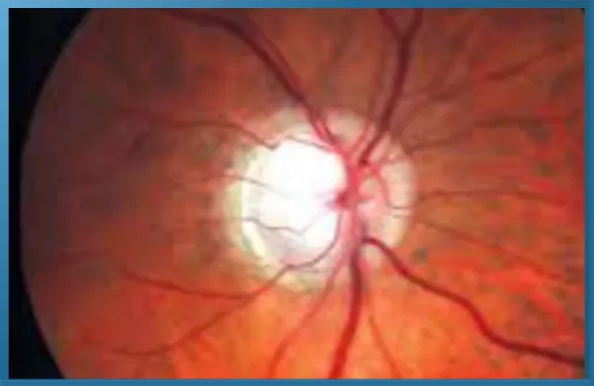
MAIN SYMPTOMS OF PRIMARY OPEN-ANGLE
GLAUCOMA
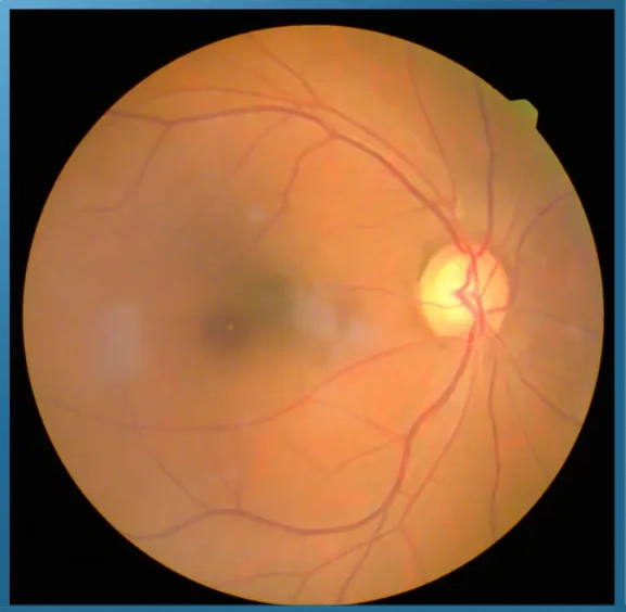
Normal-tension glaucoma is a type of glaucoma that develops even when intraocular pressure (IOP) is within the normal range. While poor aqueous humor flow was previously thought to be the main cause of glaucoma, leading to increased IOP and compression/damage of the optic nerve, in Japan, normal-tension glaucoma, where IOP is within the normal range but the optic nerve is damaged, accounts for 70% of all cases, rather than high-IOP glaucoma. Because IOP is within the normal range, it was often overlooked in previous eye examinations, but it can be detected by using fundus examination and visual field testing in combination.
The mechanism of onset is not clearly understood, but it is thought to be caused by factors such as the optic nerve’s low tolerance to IOP and poor blood circulation to the optic nerve, and genetic factors are also believed to play a role. Normal-tension glaucoma is characterized by its slow progression of symptoms, and because subjective symptoms such as pain do not appear until it worsens, it is often difficult to notice, and by the time symptoms are noticed, it may have already progressed. Furthermore, even if the field of vision in only one eye is narrowed, people often don’t notice the disease if they are living with both eyes. The only way to detect normal-tension glaucoma early is to undergo regular eye examinations.
NORMAL-TENSION GLAUCOMA IS MORE LIKELY TO
OCCUR IN THE FOLLOWING TYPES OF PEOPLE:
- Individuals with severe myopia (unable to see
the top line of the eye chart without glasses) - Individuals with a close relative who has
glaucoma - Individuals with low blood pressure
- Individuals who frequently experience
headaches - Individuals who are prone to feeling cold
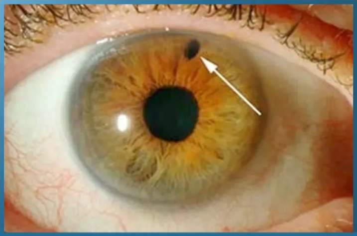
Secondary glaucoma is a type of glaucoma that develops due to elevated intraocular pressure caused by injury, illness, or side effects of medications. Glaucoma caused by injury is called “traumatic glaucoma,” and occurs when the eye is severely impacted during sports, combat sports, or accidents, damaging the angle of the eye and causing increased intraocular pressure, leading to glaucoma. Even if there are no noticeable symptoms immediately after the injury, glaucoma may develop several years later.
Next, there are several types of glaucoma caused by disease. Diabetic retinopathy, caused by diabetes, can lead to bleeding due to blockage of blood vessels in the retina, and as the condition worsens, new blood vessels proliferate to compensate for the damaged capillaries. If these new blood vessels block the angle of the eye, it causes “neovascular glaucoma.” Other types of glaucoma include “lenticular glaucoma,” which occurs when the lens expands due to cataracts and blocks the angle of the eye; “Posner-Schlossmann syndrome,” caused by uveitis; and “hemolytic glaucoma,” which occurs when bleeding in the eye obstructs the flow of aqueous humor. Glaucoma caused by the effects of medications is also called “steroid glaucoma” because it develops when the drainage of aqueous humor becomes difficult due to the side effects of steroids.
However, before discontinuing steroid administration, it is necessary to consult thoroughly with the attending physician about the condition of the disease being treated. In the case of secondary glaucoma, if the underlying disease improves, it can be expected that the progression will stop in most cases.
INDIVIDUALS WITH THESE CHARACTERISTICS ARE AT
RISK OF DEVELOPING SECONDARY GLAUCOMA:
- Individuals who have suffered a severe blow
to the eye - Individuals diagnosed with diabetes
- Individuals diagnosed with uveitis
- Individuals diagnosed with retinal disease
- Individuals receiving steroid medication
EYE SOS SIGNALS INDICATING GLAUCOMA
Glaucoma is an eye disease that affects the optic nerve. People are especially susceptible to eye diseases such as glaucoma and cataracts after the age of 40. If you notice even the slightest abnormality in your eyes, it’s crucial to see an ophthalmologist and get examined as soon as possible for early detection. Common symptoms of eye problems include eye strain and blurred vision, but if these symptoms persist even after resting your eyes, an underlying disease should be suspected. While you might think, “I’m just a little tired,” an eye disease may actually be progressing. Glaucoma, in particular, can lead to blindness if left untreated, so you should see an ophthalmologist promptly if any of the following occur.
- While eye strain is the most common cause, persistent symptoms may indicate a disease affecting the cornea, iris, lens, vitreous humor, retina, or optic nerve.

- These symptoms may be difficult to notice when using both eyes, but they can sometimes be detected when using only one eye. A narrowing of the field of vision is called “visual field constriction,” while the presence of blind spots in the field of vision may indicate “visual field defect.” These symptoms can be caused by diseases such as stroke or brain tumors, or they can be related to retinal diseases such as glaucoma or retinal detachment.

- Iridescence is a condition where a rainbow appears around a light source, such as a fluorescent lamp. Iridescence can be caused by problems with the cornea or lens, but it can also occur if glaucoma causes corneal opacity.
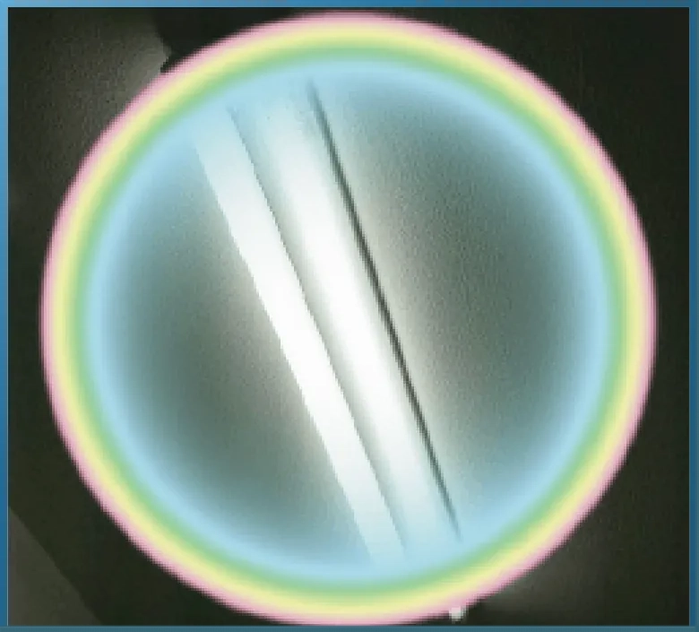
- Sudden eye pain may be caused by a rapid increase in intraocular pressure. If accompanied by severe headache or nausea, acute glaucoma should be suspected. Even if you haven’t felt anything unusual the day before, symptoms can appear suddenly, so you need to see an ophthalmologist immediately. In cases of acute glaucoma, failure to receive appropriate treatment within 48 hours can lead to blindness.
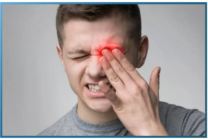
There are several types of glaucoma, but with the exception of normal-tension glaucoma, it is a disease in which the optic nerve is damaged due to elevated intraocular pressure, leading to vision loss. Damaged optic nerves and lost vision cannot be restored. The main goal of glaucoma treatment is to control elevated intraocular pressure and halt the progression of the disease as much as possible.
TREATMENT OF GLAUCOMA WITH MEDICATION
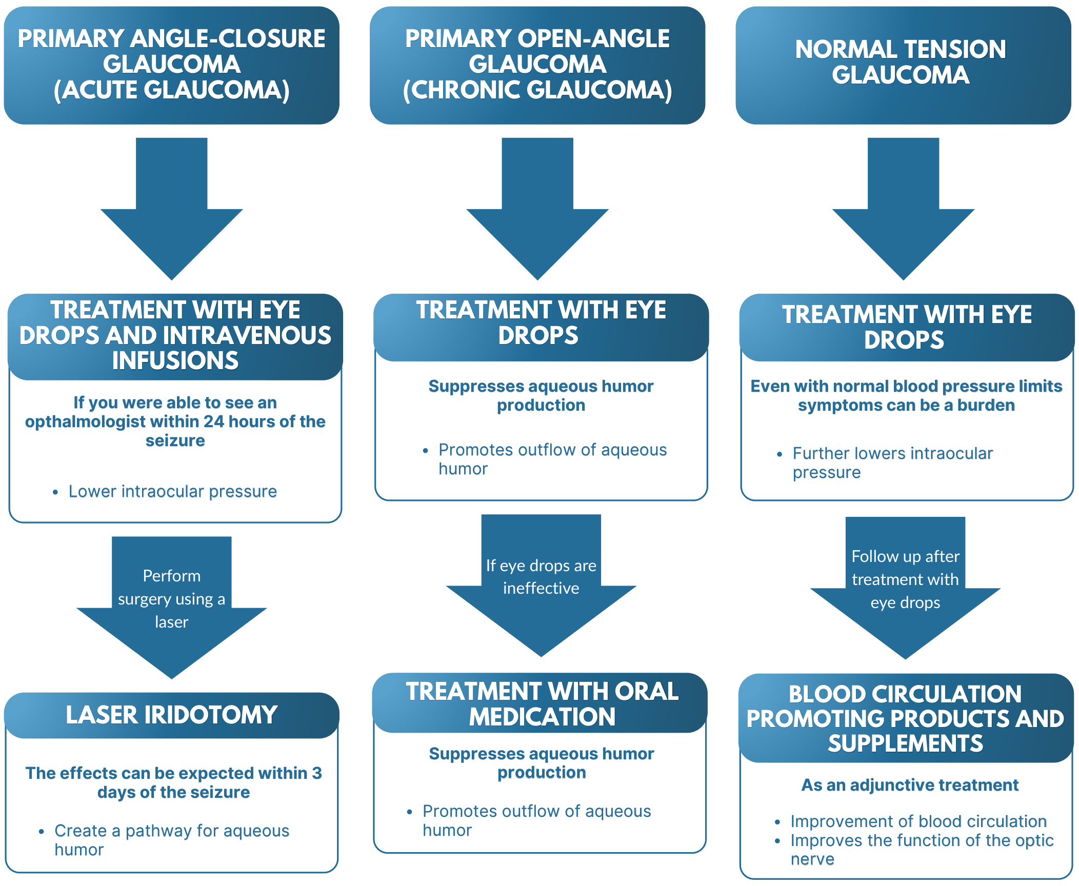
TREATMENT OF GLAUCOMA WITH EYE DROP
Eye drops used to treat glaucoma are mainly divided into three types. Treatment begins with one type of eye drop to lower elevated intraocular pressure, and while observing its effect, it may be combined with other eye drops or the medication may be changed.
TREATMENT DRUGS THAT SUPPRESS AQUEOUS HUMOR
PRODUCTION
- The sympathetic nervous system is responsible for keeping the body active. By stimulating this sympathetic nervous system, the production of aqueous humor is suppressed.
- By binding to beta-receptors in the autonomic nervous system, they suppress the function of the sympathetic nervous system and reduce the production of aqueous humor.
MEDICATIONS THAT PROMOTE AQUEOUS HUMOR
OUTFLOW
- These drugs work on the parasympathetic nervous system, which calms the body, and promote the drainage of aqueous humor.
- Prostaglandins are substances in the body that promote the excretion of aqueous humor.
A THERAPEUTIC DRUG WITH TWO EFFECTS: SUPPRESSING
AQUEOUS HUMOR PRODUCTION AND PROMOTING ITS DRAINAGE.
- Alpha-blockers work by contracting the ciliary muscle, thereby promoting the drainage of aqueous humor, while beta-blockers work by suppressing the production of aqueous humor secreted from the ciliary body.
Because eye drops used to treat glaucoma are potent, it is crucial to strictly follow the prescribed dosage and frequency. Furthermore, it is extremely dangerous to stop using the eye drops or change the dosage or frequency based on self-diagnosis, such as “the eye pain has disappeared” or “my intraocular pressure has decreased.” Stopping intraocular pressure control will inevitably lead to the progression of glaucoma, resulting in visual field defects and constriction. Using prescribed eye drops as directed and monitoring your condition is essential for effective glaucoma treatment.
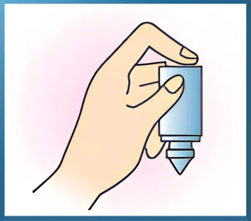
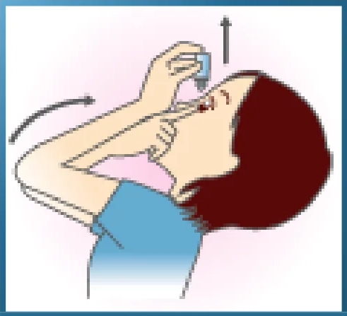
1. HOLD THE EYE DROPS IN YOUR DOMINANT HAND
AND ADMINISTER THEM.
- Hold the eye drop container between your thumb and middle finger, and press down on the bottom with your index finger.
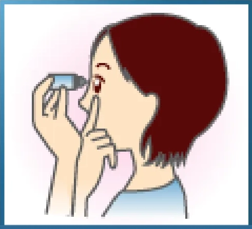
2. USE THE INDEX FINGER OF YOUR OTHER
HAND TO PULL YOUR LOWER EYELID
DOWNWARDS.
- Pull the lower eyelid of the eye you are going to instill the eye drops into downwards.
3. LOOK STRAIGHT UP AND DROP ONE DROP OF
EYE DROPS INTO YOUR EYE.
- One drop of eye drops is usually sufficient. Using more than necessary can easily lead to side effects.
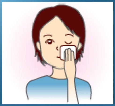
4.AFTER APPLYING THE EYE DROPS, CLOSE
YOUR EYES AND GENTLY PRESS THE INNER
CORNER OF YOUR EYE FOR ABOUT TWO
MINUTES.
- Immediately after applying the eye drops, blinking will cause the medication to be washed away by tears, so close your eyes and gently press near the inner corner of your eye to allow the eye drops to spread evenly.
*** Caution ***
*If eye drops get on your skin, wipe them off with a tissue. The medication may cause skin discoloration.
*When using multiple eye drops, leave at least 5 minutes between applications.
*Be careful not to let the tip of the eye drop container touch your eye directly.
TREATMENT OF GLAUCOMA WITH
ORAL MEDICATION
If eye drops are ineffective, oral medication will be used in combination. The oral medications used to treat glaucoma are called “carbonic anhydrase inhibitors,” and they have the effect of suppressing the production of aqueous humor.
Oral medications are considered to be more effective than eye drops, but they can have side effects, so there are restrictions on the amount and duration of administration. If no effect is seen after taking the medication for a certain period, surgery will be performed.
• Rash • Numbness in hands and feet • Fever • Nausea •
Loss of appetite • Frequent urination • Kidney stones
SURGICAL TREATMENT FOR
GLAUCOMA
Glaucoma is treated starting with medication to control elevated intraocular pressure. However, if eye drops or oral medications are not effective, surgery is chosen. In the case of angle-closure glaucoma, the angle that drains aqueous humor is blocked, so surgery is chosen from the outset in about 90% of cases.
There are two types of surgery: surgical surgery using a scalpel and laser surgery. Laser surgery is less burdensome for the patient, but if laser surgery is ineffective, surgical surgery is chosen.
LASER SURGERY FOR GLAUCOMA
Laser surgery is performed under topical anesthesia. The procedure takes only about 10 minutes, so it is minimally invasive and you can go home on the same day.
IRIDOTOMY
This surgery is performed for primary angle-closure glaucoma. In primary angle-closure glaucoma, the iris and lens are fused together, blocking the passage of aqueous humor. Therefore, a small hole of about 1 mm is made in the iris with a laser beam to create a new bypass for the aqueous humor to pass through. However, irisotomy is a race against time, and the limit for expecting results is within 3 days of the onset of the disease. After 3 days, the iris and lens will adhere together, and the surgery will not be effective. Also, while primary angle-closure glaucoma often occurs in only one eye, there is a risk of it occurring in the other eye as well, so the procedure is performed on both eyes as a preventative measure. This surgery can cause complications such as iritis and corneal opacity. In addition, the surgery may not be possible if the distance between the cornea and iris is too close.
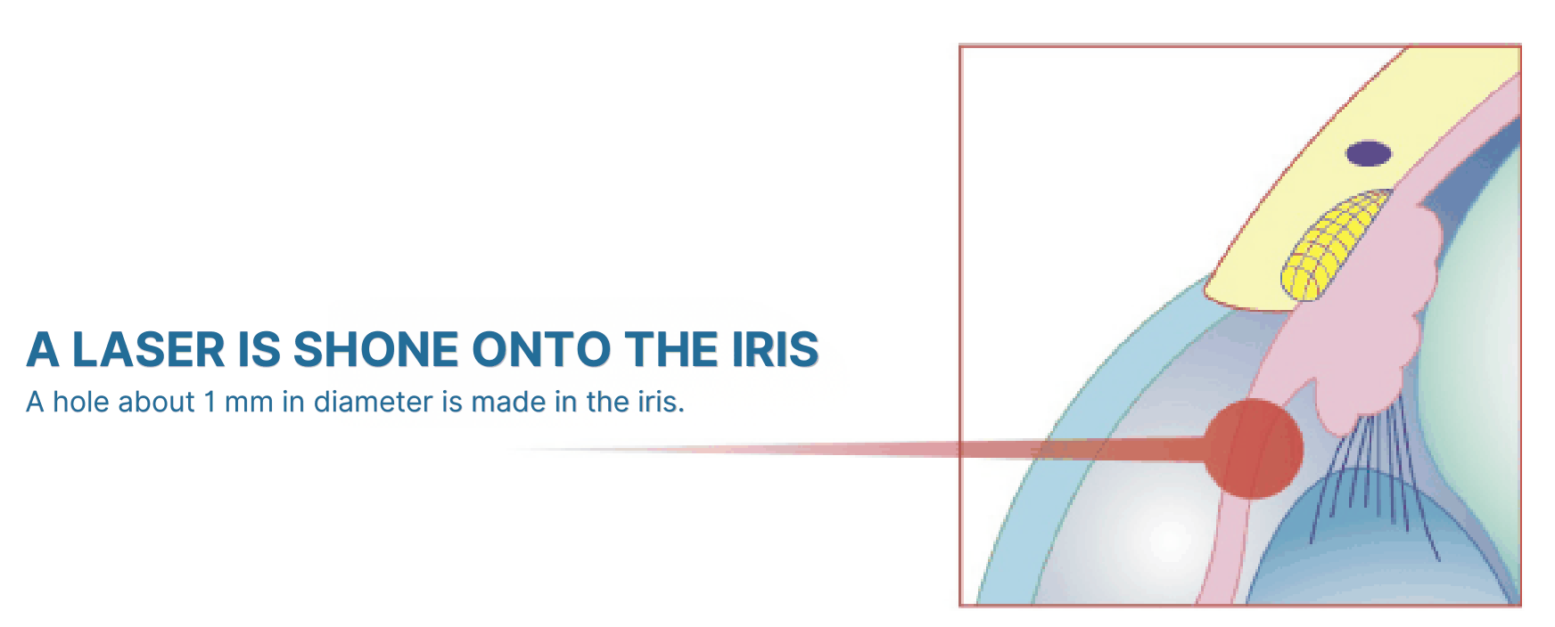
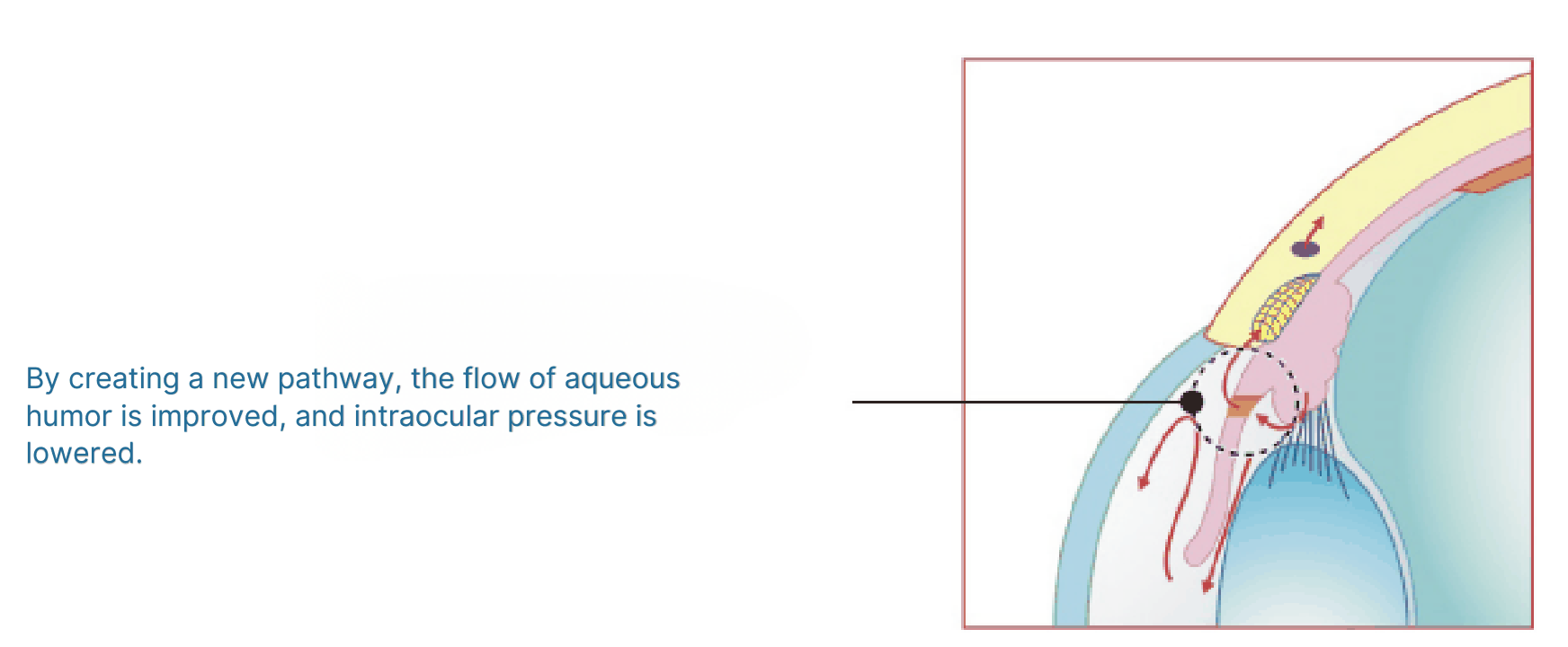
LASER IRIDOTOMY CAN LOWER INTRAOCULAR PRESSURE AND
SUPPRESS THE PROGRESSION OF GLAUCOMA.
TRABECULOPLASTY
This surgery is performed for primary open-angle glaucoma. Anterior to Schlemm’s canal, the drainage outlet for aqueous humor, there is a reticular tissue called the “trabecular meshwork” that acts as a filter. Primary open-angle glaucoma develops when the trabecular meshwork becomes blocked, so this surgery uses a laser to improve the flow of aqueous humor through the trabecular meshwork. The surgery takes about 5 to 10 minutes and has almost no side effects. The effect may wear off after about 1 to 2 years, in which case a second surgery will be performed.
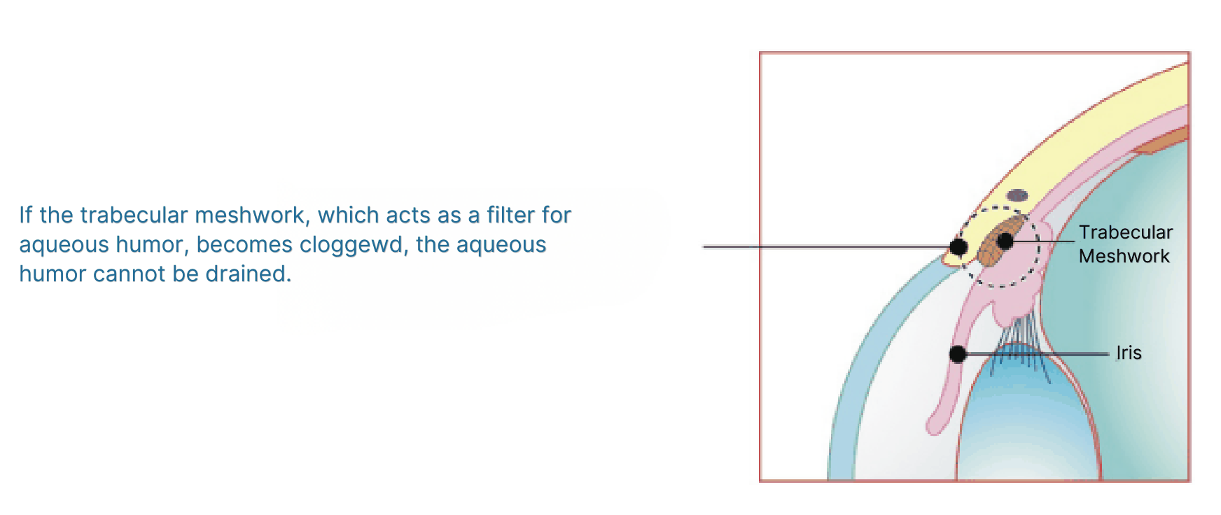
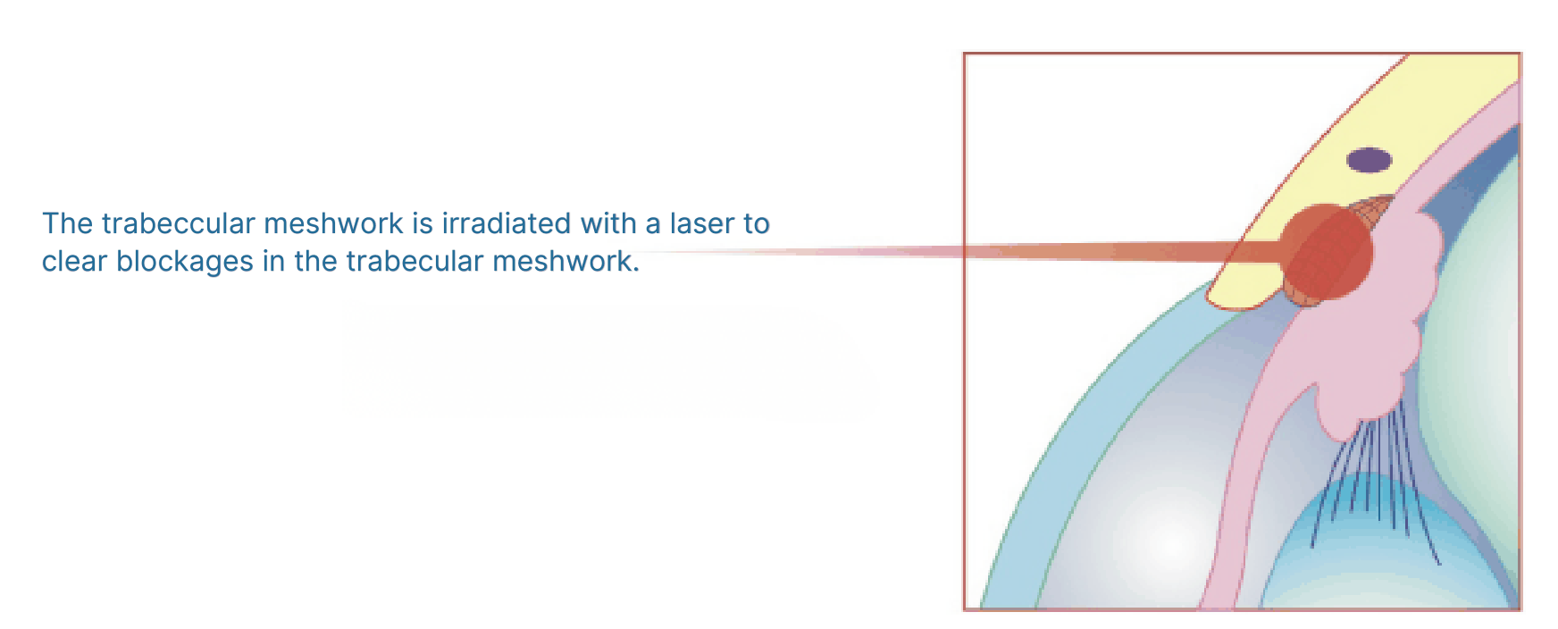
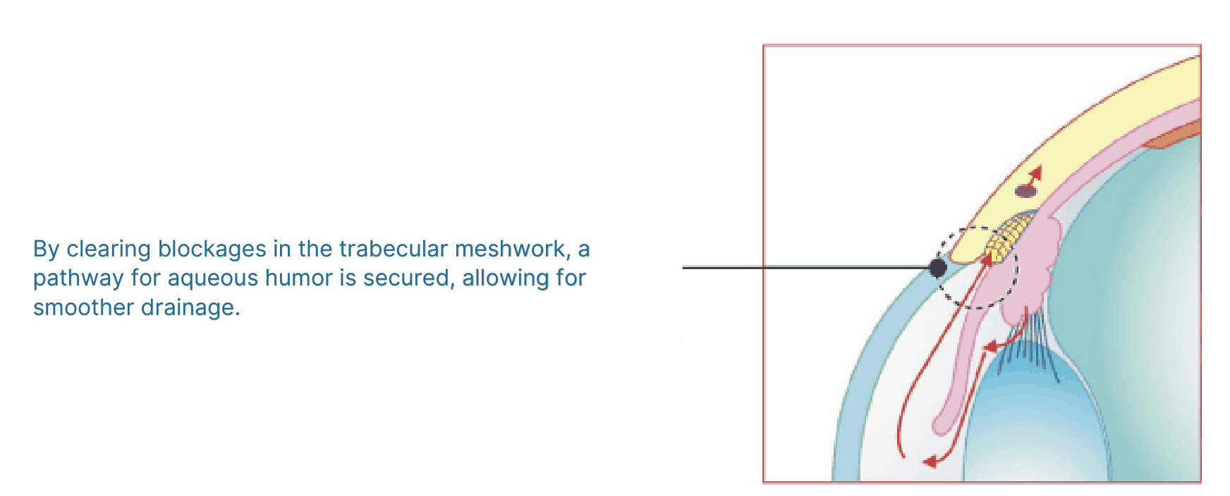
LASER TRABECULOPLASTY CAN LOWER INTRAOCULAR PRESSURE AND
SUPPRESS THE PROGRESSION OF GLAUCOMA.
SURGICAL TREATMENT FOR GLAUCOMA
Surgical procedures are burdensome for patients, but if drug therapy or laser surgery is ineffective, surgical procedures using a scalpel are performed. There are several types of surgical procedures.
TRABECULECTOMY
A portion of the sclera is removed to create a new outlet for aqueous humor.
TRABECULAR INCISION
We cut open the blocked sections of the trabecular meshwork to improve the flow of aqueous humor.
GONIOSYNOLYSIS
This treatment separates the adhesions between the iris and the angle of the iridocorneal cavity, improving the flow of aqueous humor.
CILIARY BODY DESTRUCTION
It destroys the ciliary body that secretes aqueous humor, thereby reducing the amount of aqueous humor produced.
IMPLANT SURGERY
A device to absorb aqueous humor and an artificial tube are attached to create a pathway for the aqueous humor to flow through.
Glaucoma is the leading cause of blindness in Japan. While it was once said that “glaucoma equals blindness,” advances in medicine have allowed most people to maintain their field of vision and visual acuity.
The most important things for glaucoma are early detection, early treatment, and regular checkups with an ophthalmologist to continue treatment. When subjective symptoms such as eye pain improve, awareness of treatment may diminish.
Stopping eye drops or changing the frequency or amount of eye drops on your own can worsen the disease, so it is crucial to follow your doctor’s instructions and continue regular checkups.
After the age of 40, various eye diseases, not just glaucoma, become more common. The eyes are vital organs that receive 80% of the information we get from the outside world. To protect your precious eyes, having a “primary eye doctor” you can consult at any time can lead to the early detection of diseases.